FONA is a common term and is used in this session to mean a 'Front of Neck Airway'.
FONA can mean a variety of things, in terms of procedure and urgency. All refer to an artificial airway formed in the front of the patient's neck. All involve making a path from the skin through the soft tissues of the anterior neck to the trachea. The trachea is accessed and a tube is inserted:
Site
The site for the FONA is usually at the cricothyroid membrane (Fig 1). It can be lower in the neck between the second and third tracheal rings, similar to a formal tracheostomy.
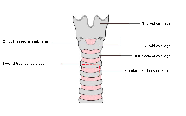
Fig 1 Cricothyroid membrane
Urgency
In an emergency, a cricothyroidotomy is almost always quicker and easier to perform than a tracheostomy.
When working as part of a team managing a situation where FONA is required, it is vitally important to communicate clearly.
Asking a surgeon to 'perform FONA' could be interpreted as 'please perform a tracheostomy'. In an emergency, a surgical cricothyroidotomy is almost always the fastest method for FONA.
This potential problem is discussed in a joint editorial from the Royal College of Anaesthetists, the Royal College of Surgeons, ENT UK and the Difficult Airway Society [1] (read details regarding the reference).
Incision
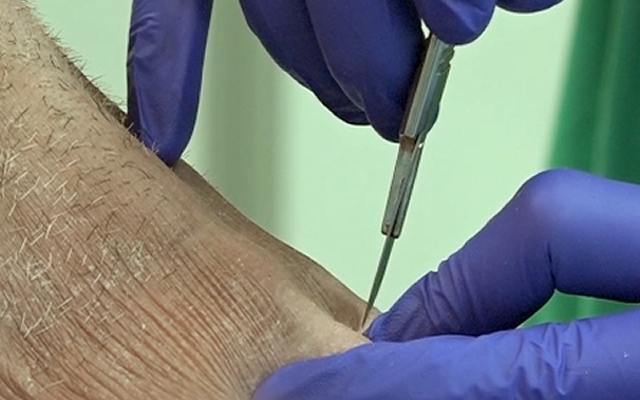
The incision can be made with a scalpel blade or percutaneously by the Seldinger technique, using a needle/wire/dilator (Fig 2).
Access can be achieved with a needle alone, but these devices can be unreliable and require a high pressure gas source for ventilation.
Some bespoke devices are available that use variations on these two basic techniques.
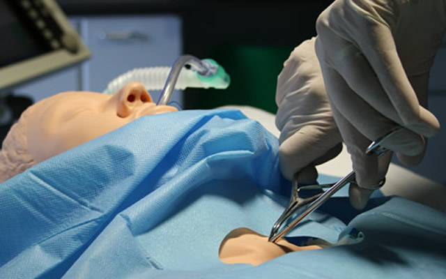
Fig 2 Seldinger technique
Larger tracheostomy stoma
A larger tracheostomy stoma usually needs a size 7-9 mm tracheostomy tube (Fig 4).
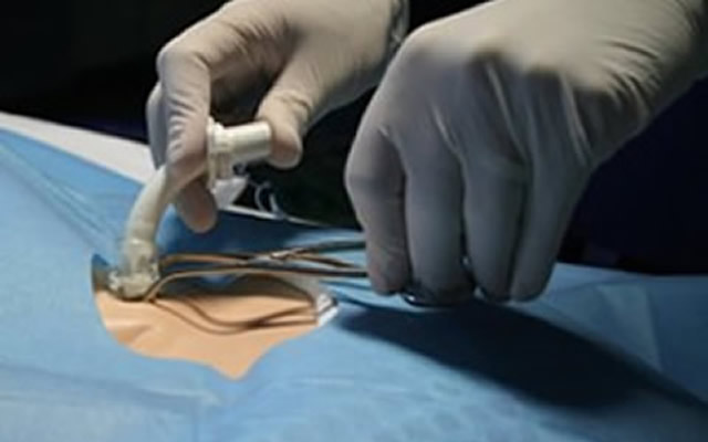
Fig 4 Larger tracheostomy stoma
Cricothyroidotomy
For a cricothyroidotomy, a 6 mm internal diameter tube is usually used in adults (Fig 3). Specific cricothyroidotomy tubes are available, although a size 6.0 endotracheal tube also works well. Smaller tubes may be easier to insert but only provide emergency oxygenation. They are too small to allow effective ventilation and removal of carbon dioxide. Tubes with a cuff are a little more bulky to insert but offer a degree of protection against aspiration and allow effective positive pressure ventilation.
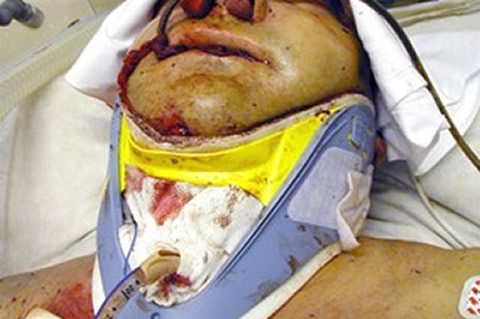
Fig 3 Cricothyroidotomy
Fig 1 shows a smaller, cuffed cricothyroidotomy tube through the cricothyroid membrane.
Fig 2 shows a larger cuffed tracheostomy tube, sited lower in the neck.
FONA is a common term and is used in this session to mean a 'Front of Neck Airway'.
FONA can mean a variety of things, in terms of procedure and urgency. All refer to an artificial airway formed in the front of the patient's neck. All involve making a path from the skin through the soft tissues of the anterior neck to the trachea. The trachea is accessed and a tube is inserted.
- Site
- Urgency
- Incision
- Once the stoma has been created, an artificial airway device is
required to keep it patent:
- Cricothyroidotomy
- Larger tracheostomy stoma
- Cricothyroidotomy vs tracheostomy
- Different types of tube:
Cricothyroidotomy vs tracheostomy
Fig 5a shows a smaller, cuffed cricothyroidotomy tube through the cricothyroid membrane.
Fig 5b shows a larger cuffed tracheostomy tube, sited lower in the neck.
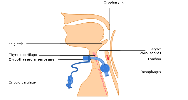
Fig 5a Smaller cuffed cricothyroidotomy tube
FONA is a common term and is used in this session to mean a 'Front of Neck Airway'.
FONA can mean a variety of things, in terms of procedure and urgency. All refer to an artificial airway formed in the front of the patient's neck. All involve making a path from the skin through the soft tissues of the anterior neck to the trachea. The trachea is accessed and a tube is inserted.
- Site
- Urgency
- Incision
- Once the stoma has been created, an artificial airway device is
required to keep it patent:
- Cricothyroidotomy
- Larger tracheostomy stoma
- Cricothyroidotomy vs tracheostomy
- Different types of tube:
Cricothyroidotomy vs tracheostomy
Fig 5a shows a smaller, cuffed cricothyroidotomy tube through the cricothyroid membrane.
Fig 5b shows a larger cuffed tracheostomy tube, sited lower in the neck.
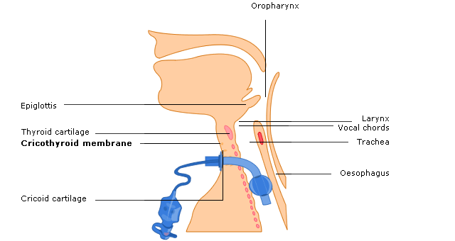
Fig 5b Larger cuffed tracheostomy tube
Uncuffed
Fig 6 shows an inserted uncuffed tube. If the upper airway is patent, air can flow through the mouth and nose, through the tracheostomy tube and past the tube in the trachea.
However, because there is no seal in the trachea, the airway is not protected against aspiration and positive pressure ventilation cannot be effectively applied via the tube.

Fig 6 Uncuffed tube
Cuffed
Fig 7 shows a cuffed tracheostomy tube. The cuff seals off the upper airway. When the tube is correctly positioned within the trachea, gas can only move into and out of the lungs via the tracheostomy tube.
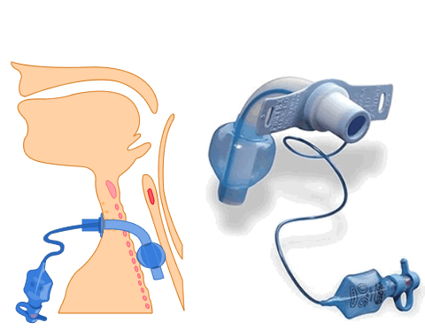
Fig 7 Cuffed tube