Tracheostomy may be temporary or long term/permanent, and may be formed electively or as an emergency procedure. They may also be classified by their method of initial insertion – either surgical or percutaneous.
Most experts agree that within a week or two following insertion, it doesn’t matter too much how the tracheostomy was originally inserted. However, long term follow up studies are lacking.
Unless a particular approach is clearly indicated, for most patients, a team approach is recommended with discussion between ICU and surgical colleagues about the best option for the patient.
Temporary tracheostomies
Temporary tracheostomies are required for ICU patients who are weaning from invasive ventilatory support or when there is a temporary problem with the upper airway (obstruction, aspiration risk, swallowing problem or for secretion management) These tubes will be removed when the patient recovers.
Long term/permanent tracheostomies
Some patients need chronic respiratory support, or they have a long term or irreversible upper airway problem. This requires a permanent tracheostomy. Examples include severe or progressive neurological conditions or patients with insufficient respiratory capacity to breathe without support.
If the larynx is surgically removed during a laryngectomy, the stoma created is an end-stoma (the trachea now ends at the front of the neck). This is irreversible and the patient is a neck-only breather.
Surgical tracheostomies
Surgical tracheostomy is usually carried out in an operating theatre where conditions are sterile, and lighting is good. General anaesthesia is most frequently used however this technique can also be carried out with a local anaesthetic.
A surgical opening is made into the trachea into which a tube is placed; this may then be sutured to the skin or secured with cloth ties or a holder.
Because a stoma has been cut and stitched, surgical tracheostomies become established tracts (from skin to trachea) within around 2-3 days. A percutaneous tracheostomy dilates and stretches the tissues which will spring back if a tube is removed. A percutaneous tracheostomy should ideally not be changed for 7-10 days. This is one of the reasons why the type of tracheostomy performed is essential information on the bedhead sign if the tracheostomy tube becomes displaced.
Percutaneous tracheostomies
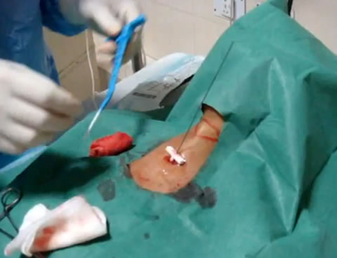
Percutaneous tracheostomy the most commonly used technique in critical care as it is simple and quick, and can be performed by non-surgeons, following appropriate training. One advantage of a percutaneous procedure is that it can be performed at the bedside of a patient in the ICU, removing the need to transfer potentially unstable patients and the problems in theatre scheduling.
The procedure involves the insertion of a needle through the neck into the trachea followed by a guide-wire through the needle. The needle is removed and the tract made gradually larger by inserting a series of progressively larger dilators over the wire until the stoma is large enough to fit a suitable tube (Seldinger technique). This is then secured by cloth ties or a holder.
The session resources, accessed via the Resources button, contains links to three videos of percutaneous tracheostomy insertion.
Bleeding risks
Generally, a surgical approach is favoured in children, if the anatomy is difficult or significant blood vessels are in the way of the stoma site. For patients with abnormal clotting, a percutaneously formed stoma may create less trauma and the tube may tamponade any oozing from the wound. However, a surgical approach may allow more direct control of bleeding along with access to diathermy. Hybrid approaches have been described.