A Tracheostomy Tube <i>in situ</i>
The structures you have identified are shown on this slide in cross section to demonstrate the path of the tracheostomy stoma and any tubes or devices inserted through it.
Fig 1 shows a sagittal plane image of the relevant anatomy in cross section. Fig 2 shows where the tracheostomy tube sits when in situ.
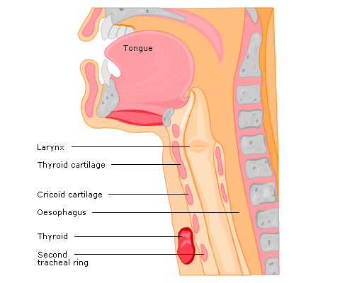
Fig 1 Sagittal plane image showing the relevant anatomy in cross section
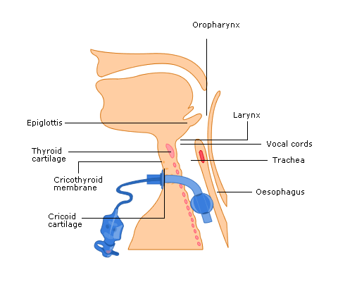
Fig 2 A cuffed tracheostomy tube had been inserted through the anterior neck and the tube tip is sitting correctly in the trachea